
-
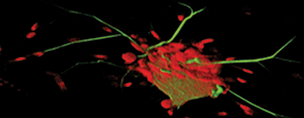 3D image of sensory neuron and surrounding satellite cells (Twiss lab - Dr. E van Niekerk)
 -
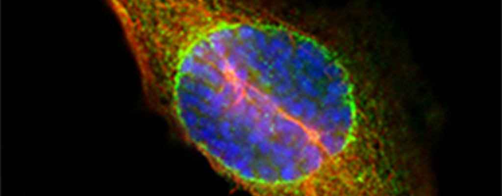 Prophase nuclear envelope invagination with Lis1 protein in green (Smith lab)
 -
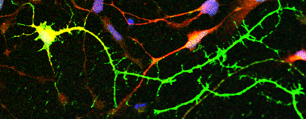 Human cortical neuron derived from induced pluripotent stem cells (Lizarraga lab)
 -
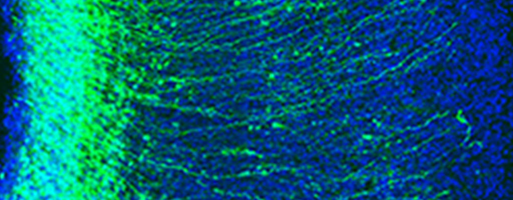 Image of mouse brain with GFP expressing cortical neurons (Lizarraga lab)
 -
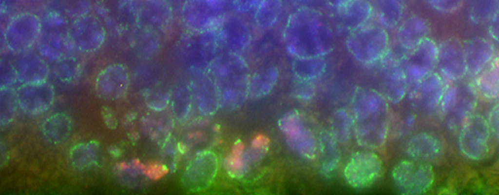 Prophase nuclear envelope invagination in E13.5 subventricular zone stem cells (Smith lab)
 -
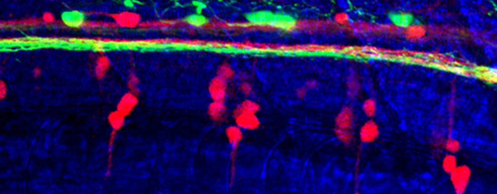 Zebrafish spinal cord (Poulain lab)
Research programs in the Center for Childhood Neurotherapeutics are broadly focused on neural connectivity. The human brain contains billions of neurons, and connections between these neurons, termed synapses, make the brain and spinal cord work. Neurons extend long cytoplasmic processes, ‘dendrites’ and ‘axons’, to form these connections with other neurons and with their target tissues (e.g., muscle). In many vertebrate organisms including humans, the axons can extend for a meter or more distance. Developing and maintaining such long cellular extensions is a daunting, but essential undertaking. Injury to axons through disease or trauma presents new challenges if any function is to be recovered. Research projects in the Center are focused on how development, function, and repair of neural connectivity.
Funding for work in the Center has included awards from the National Institutes of Health, National Science Foundation, the US Department of Defense, the Dr. Miriam and Sheldon G. Adelson Medical Research Foundation, and the US-Israel Binational Science Foundation. The Center Faculty pages outline research projects of Drs. Lizarraga, Poulain, Smith and Twiss in more detail.